
Primary areas of research
Functionalization of small-molecules as phototheranostic agents
Small molecules as phototheranostics are uniquely positioned for advancing the field of current therapeutic monitoring. This involves a single, well-defined chemical structure that upon NIR irradiation, generates both optoacoustic contrast for imaging (diagnostic) and exerts an ROS-mediated therapeutic effect at the same site without the batch-to-batch variability, synthetic complexity, or clearance concerns of nanoformulations. We design and synthesize NIR-active small-molecule agents such as asymmetric, glycerol-substituted zinc phthalocyanines that pair deep-tissue photoacoustic readout with photodynamic therapy, achieving long circulation, tumor accumulation, and activity even in hypoxic, treatment-resistant tumors without added encapsulation.
We extend the same molecular-design logic to developing targeted theranostics with third generation photosensitizer modifications.

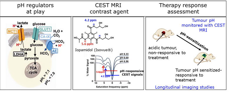
Imaging extracellular tumor pH to guide therapeutic outcomes
Extracellular acidosis and reprogrammed metabolism are hallmarks of aggressive solid tumors, and they actively blunt chemo-, radio-, and immunotherapy. Using acidoCEST (acido-Chemical Exchange Saturation Transfer) MRI, pH-responsive contrast agents, and metabolically targeted photoacoustic probes, we noninvasively map these features and test whether correcting them restores treatment sensitivity. In other studies, we have shown that tumor acidosis drives immune escape, and that a pH-sensitizer can potentiate immune checkpoint blockade pointing toward imaging-defined windows for more effective combination therapy.
Our group currently focuses on developing a novel regimen of pH neutralization for epithelial ovarian cancer and aims to demonstrate improved efficacy of first line chemotherapy drugs in this model with the proposed pH-neutralization. Finally, for studying the in vivo validation of this improved therapy response, we focus on CEST MRI performed on a 3T Siemens Prisma MR scanner with a pH-responsive CEST MRI contrast agent.
Metabolic imaging and magnetic resonance spectroscopy (MRS) of cancer
Altered metabolism is a central hallmark of cancer, and reading it out noninvasively can reveal disease and therapy response earlier than anatomy alone. We probe tumor metabolism with complementary tools: targeted photoacoustic contrast agents that report metabolic activity in living tumors, and proton magnetic resonance spectroscopy (¹H-MRS) to quantify metabolite profiles in ovarian cancer. Using preclinical models of metastatic ovarian cancer, we have shown that the lactate/lipid signal- a marker of glycolytic activity drops markedly after chemotherapy while choline persists, demonstrating that MRS can noninvasively capture biochemical signatures of treatment response (ISMRM 2026). By tracking how these metabolic readouts shift across disease progression and therapy, we aim to define imaging biomarkers that enable earlier detection, stratify aggressive disease, and guide treatment decisions in gynecologic and other cancers.
